DCTD
PDB:2W4L
Revision
Revision Type:created
Revised by:created
Revision Date:created
Entry Clone Accession:BC001286
Entry Clone Source:Mammalian Gene Collection
SGC Clone Accession:Tag:C-terminal hexahistidine tag: -ahhhhhh
Host:E.coli BL21(DE3) R3 pRARE, where R3 denotes a derivative of BL21(DE3) resistant to a strain of T1 bacteriophage (SGC Oxford) and the pRARE plasmid originating from the Rosetta strain (Novagen) supplies tRNAs for rare codons.
Construct
Prelude:Sequence:MSCKKRDDYLEWPEYFMAVAFLSAQRSKDPNSQVGACIVNSENKIVGIGYNGMPNGCSDDVLPWRRTAENKLDTKYPYVCHAELNAIMNKNLTDVKGCSMYVALFPCNECAKLIIQAGIKEVIFMSDKYHDSDEATAARLLFNMAGVTFRKFIPKCSKIVIDFDSINSRPSahhhhhh
Vector:pNIC-CH2
Growth
Medium:Antibiotics:Procedure:Cells from a glycerol stock were grown in 20 ml TB supplemented with 8 g/l glycerol, 100 µg/ml kanamycin and 34 µg/ml chloramphenicol at 30 °C overnight. The overnight culture (10 ml) was used to inoculate 0.75 l TB supplemented with 8 g/l glycerol, 50 µg/ml kanamycin and approximately 0.4 ml 204 Antifoam A6426 (Sigma). The culture was grown in a LEX bioreactor system (Harbinger Biotechnology) at 37 °C until OD600 reached ~2. The bottle was down-tempered to 18 °C over a period of 1 hour before target expression was induced by addition of 0.5 mM IPTG. Expression was allowed to continue overnight and cells were harvested the following morning by centrifugation (4,400 x
g, 10 min, 4 °C). The resulting cell pellet (21 g wet cell weight) was resuspended in lysis buffer (1.5 ml/g cell pellet), supplemented with 500 U Benzonase (Merck) and 0.5 tablet of Complete EDTA-free protease inhibitor (Roche Applied Science). The cell suspension was stored at -80 °C.
Purification
ProcedureColumnsIMAC: Ni-charged 1 ml HiTrap Chelating HP (GE Healthcare)
Gel filtration column: HiLoad 16/60 Superdex 75 Prep Grade (GE Healthcare)
Procedure
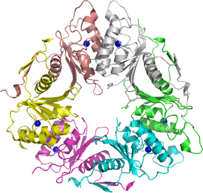
Purification of the protein was performed as a two step process on an ÄKTAxpress system (GE Healthcare). Prior to purification, columns were equilibrated with IMAC wash1 buffer and gel filtration buffer, respectively. The filtered lysate was loaded onto the Ni-charged HiTrap Chelating column and washed with IMAC wash1 buffer followed by IMAC wash2 buffer. Bound protein was eluted from the IMAC column with IMAC elution buffer and automatically loaded onto the gel filtration column. The target protein was eluted as a single peak from the gel filtration column but appeared to be larger than a tetramer. Later, the crystal structure confirmed a homohexamer. Fractions containing the target protein were pooled and fresh TCEP was added to a final concentration of 2 mM. The protein was subsequently concentrated using an Amicon centrifugal filter device with 10,000 NMWL (Millipore) to 12.1 mg/ml in a volume of 1.6 ml.
Extraction
ProcedureThe cell suspension was quickly thawed in water. Cells were disrupted by sonication (Vibra-Cell, Sonics) at 80% amplitude for 3 min effective time (pulsed 4s on, 4s off) and cell debris was removed by centrifugation (49,100 x
g, 20 min, 4 ºC). The supernatant was decanted and filtered through a 0.45 µm flask filter.
Concentration:LigandMassSpec:Crystallization:Crystals were obtained by the hanging drop vapour diffusion method in a 24-well plate containing 500 µl well solution. 1 µl of the protein solution (12.1 mg/ml) was mixed with 1 µl of well solution consisting of 0.2 M sodium thiocyanate and 6% PEG 3350. The plate was incubated at room temperature and crystals appeared within 24 hours. However, the crystallization condition was not stable and precipitated initially. The crystals were quickly transferred to cryo solution containing 0.2 M sodium thiocyanate, 0.3 M NaCl, 8% PEG 3350 and 25% glycerol and flash frozen in liquid nitrogen.
NMR Spectroscopy:Data Collection:Data was collected to 2.1 Å resolution at Bessy BL14-1. The crystal belonged to space group P21 with cell parameters 66.2 80.1 96.3 90 94.3 90 and the asymmetric unit contained one hexamer.
Data Processing:The data was processed with XDS and MR and the original molecular replacement was performed using MOLREP and the CHAINSAW model of 2HVW as search model. Structure was refined with REFMAC5. Final R-values were R=20.0% and Rfree=24.3%. Coordinates and structure factors were deposited in PDB with accession code 2W4L.