CAMK2G
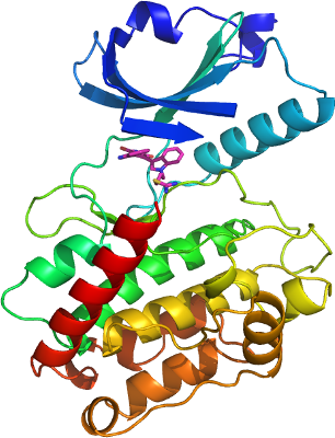
PDB:2V7O
Revision
Revision Type:created
Revised by:created
Revision Date:created
Entry Clone Accession:gi|26667203
Entry Clone Source:MGC
SGC Clone Accession:
Tag:mhhhhhhssgvdlgtenlyfq*s(m) TEV-cleavable (*) N-terminal his6 tag.
Host:BL21(DE3)-R3-pRARE2
Construct
Prelude:
Sequence:mhhhhhhssgvdlgtenlyfq*smATCTRFTDDYQLFEELGKGAFSVVRRCVKKTPTQEYAAKIINTKKLSARDHQKLEREARICRLLKHPNIVRLHDSISEEGFHYLVFDLVTGGELFEDIVAREYYSEADASHCIHQILESVNHIHQHDIVHRDLKPENLLLASKCKGAAVKLADFGLAIEVQGEQQAWFGFAGTPGYLSPEVLRKDPYGKPVDIWACGVILYILLVGYPPFWDEDQHKLYQQIKAGAYDFPSPEWDTVTPEAKNLINQMLTINPAKRITADQALKHPWVCQRSTVASMMHRQETVECLRKFNARRKLKGAILTTMLVSRNFSVG
Vector:pNIC28-Bsa4.
Growth
Medium:LB
Antibiotics:
Procedure:3ml from a 50 ml overnight culture was used to inoculate each of two flasks containing 1 litre of LB media containing 50 µg/ml kanamycin and 34 µg/ml chloramphenicol. Cultures were grown at 37°C to an OD600 of ~0.4 and then cooled to 18°C. Expression was induced for 4 hours using 0.5mM IPTG at an OD600 of 0.8. The cells were collected by centrifugation and the pellet resuspended in binding buffer and frozen.
Purification
Procedure
Column 1: Ni-affinity chromatography.
Column 2: Size exclusion chromatography (Sephacryl S200)
5 ml of 50% Ni-sepharose slurry (Amersham Biosciences) was applied to a 1.5 x 10 cm gravity column. The column was equilibrated with 50 ml binding buffer. The lysate was applied to the column which was subsequently washed with 50 ml wash buffer. CaMK2G was eluted in 5 ml step elutions containing 50 mM, 100 mM, 150 mM and 250 mM imidazole. The eluted protein was collected and analyzed by SDS-PAGE. DTT was added to the protein sample to a final concentration of 10 mM. TEV-protease was added to the protein over night. However, the N-terminal his 6 -tag was not cleaved due to phosphorylation of a serine residue within the protease cleavage site.
The cleaved protein was concentrated to 3 ml using Centricon concentrators (10kDa cut off) and applied to a Sephacryl S200 column equilibrated in SEC buffer at a flow rate of 1.0 ml/min. CaMK2G eluted at 65 minutes corresponding to a retention time of a monomeric protein of that size. Eluted fractions were 95% pure as judged by SDS-PAGE. 10 mM DTT was added to the final eluted sample.
Extraction
Procedure
Cell pellets in binding buffer plus 1 mM PMSF, 0.5 mM TCEP were lysed using a high pressure cell disrupter. The lysate was centrifuged at 17,000 rpm for 30 minutes and the supernatant collected for purification. Prior to purification the lysate was passed through a DE52 column (10g/L resin) to remove DNA.
Concentration:Centricon with a 10kDa cut off in SEC-buffer
Ligand
MassSpec:
Crystallization:The inhibitor BIM9 (AXXORA) was added to the protein in 3-fold molar excess and the complex concentrated to 13 mg/ml. Crystals were obtained in sitting drops using the vapor diffusion method by mixing 50 nl of the concentrated protein with 100 nl of a well solution containing 0.10M MgCl2, 0.1M HEPES pH 7.0, 20% PEG 6K, 10% ethylene glycol. A single crystal appeared after several days at 4°C and reached maximum size after two months.
NMR Spectroscopy:
Data Collection:The crystal was cryo-protected using the well solution supplemented with 25% ethylene glycol and flash frozen in liquid nitrogen. Diffraction data were collected at the SLS beam line X10SA to a maximum resolution of 2.25Å.
Data Processing: