BIRC1
PDB:2VM5
Revision
Revision Type:created
Revised by:created
Revision Date:created
Entry Clone Accession:gi|119393877
Entry Clone Source:In house cloning.
SGC Clone Accession:Tag:N-terminal hexahistidine tag with integrated TEV protease cleavage site: mhhhhhhssgvdlgtenlyfq*s(m)
Host: E.coli BL21(DE3) R3 pRARE, where R3 denotes a derivative of BL21(DE3) resistant to a strain of T1 bacteriophage (SGC Oxford) and the pRARE plasmid originating from the Rosetta strain (Novagen) supplies tRNAs for rare codons.
Construct
Prelude:Sequence:mhhhhhhssgvdlgtenlyfq*sMRVKNLKSRLRGGKMRYQEEEARLASFRNWPFYVQGISPCVLSEAGFVFTGKQDTVQCFSCGGCLGNWEEGDDPWKEHAKWFPKCEFLRSKKSSEEITQSYR
Vector:PNIC-Bsa4
Growth
Medium:Antibiotics:Procedure:Cells from a glycerol stock were grown in 20 ml TB supplemented with 8 g/l glycerol, 100 µg/ml kanamycin and 34 µg/ml chloramphenicol at 30 ºC overnight. The overnight culture (20 ml) was used to inoculate 1.5 l TB supplemented with 8 g/l glycerol, 50 µg/ml kanamycin and approximately 200 µl PPG P2,000 81380 anti-foam solution (Fluka). The culture was grown in a LEX bioreactor system (Harbinger Biotechnology) at 37 ºC until OD600 reached ~2. The culture was down-tempered to 18 ºC over a period of 1 hour before target expression was induced by addition of 0.5 mM IPTG. Expression was allowed to continue overnight and cells were harvested the following morning by centrifugation (4,500 x
g, 10 min, 4 ºC). The resulting cell pellet (18.4 g wet cell weight) was resuspended in lysis buffer (3 ml/g cell pellet), supplemented with 2000 U Benzonase (Merck) and one tablet of Complete EDTA-free protease inhibitor (Roche Applied Science). The cell suspension was stored at -80 ºC.
Purification
ProcedureColumnsIMAC: Ni-charged 1 ml HiTrap Chelating HP (GE Healthcare)
Gel filtration column: HiLoad 16/60 Superdex 75 Prep Grade (GE Healthcare)
Procedure
Purification of the protein was performed as a two step process on an ÄKTAxpress system (GE Healthcare). Prior to purification, columns were equilibrated with IMAC wash1 buffer and gel filtration buffer, respectively. The filtered lysate was loaded onto the Ni-charged HiTrap Chelating column and washed with IMAC wash1 buffer followed by IMAC wash2 buffer. Bound protein was eluted from the IMAC column with IMAC elution buffer and automatically loaded onto the gel filtration column. Fractions containing the target protein were pooled and fresh TCEP was added to a final concentration of 2 mM. The protein was subsequently concentrated using an Amicon Ultra-15 centrifugal filter device, 5,000 NMWL (Millipore) to 14.4 mg/ml in a volume of 1.1 ml. The identity of the protein was confirmed by mass spectrometry.
Tag removal
The N-terminal histidine tag was proteolytically removed by incubating the target protein with His-tagged TEV protease at a molar ratio of 30:1 at 4 ºC overnight. The proteolytic reaction went to completion, as judged by SDS-PAGE. For purification, a matrix mix was made by mixing equal volumes of Ni-agarose (GE Healthcare) and GF-buffer. 40 mM of imidazole was added to both sample and matrix-mix. The target protein was purified from tag and protease by adding 1 ml matrix-mix to the reaction mixture and incubating at 4ºC for two hours with stirring. The supernatant containing the cleaved protein target was recovered and concentrated using a Vivaspin centrifugal filter device with 5,000 MWCO (Sartorius). The final protein concentration was determined to 24.5 mg/ml in a volume of 0.6 ml.
Extraction
ProcedureThe cell suspension was quickly thawed in water. Cells were disrupted by sonication (Vibra-Cell, Sonics) at 80% amplitude for 3 min effective time (pulsed 4s on, 4s off) and cell debris was removed by centrifugation (49,100 x
g, 20 min, 4 ºC). The supernatant was decanted and filtered through a 0.45 µm flask filter.
Concentration:LigandMassSpec:Crystallization:Crystals were obtained by the sitting drop vapour diffusion method in a 96-well plate. 0.1 µl protein solution (diluted to 12 mg/ml) was mixed with 0.1 µl of well solution consisting of 0.1 M citric acid pH 5.0 and 20% (w/v) PEG 6000. The plate was incubated at 20 ºC and crystals appeared within a few hours. 0.5 µl cryo solution, consisting of well solution complemented with 20% glycerol and 0.3 M NaCl, was added directly to the drop and the crystal was flash frozen in liquid nitrogen.
NMR Spectroscopy:Data Collection:Data was collected at ESRF (BM-14).
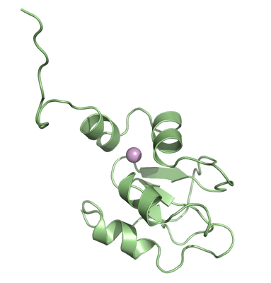
Data Processing:The crystal belonged to space group P212121 with the cell parameters 36.5, 43.1, 57.0, 90.0, 90.0, 90.0. Data was processed and scaled using XDS and XSCALE. The structure was solved by molecular replacement with MOLREP using the structure of the peptide antagonists of Melanoma Inhibitor of Apoptosis (ML-IAP) (PDB-code: 1OXN) as search model. The initial model was then automatically built using ARP/WARP. Final cycles of model building and refinement were performed in COOT and REFMAC5. Data in the interval 34 Â 1.8 Å resolution was used and at the end of the refinement the R values was: R= 17.8% and Rfree= 22%. The coordinates for the crystal structure were deposited in the Protein Data Bank, accession code 2VM5.