
| |
 |
|
 |
 |
| |
|
|
|
|
Protein tyrosine phosphatases (PTPs) play a critical role regulating cell signaling by selectively dephosphorylating their substrates. So far our laboratory has released 22 human high resolution crystal structures which, together with prior structural knowledge, results in an excellent structural coverage of the classical PTP family. A recent structural analysis carried out in our laboratory compared the existing structural information available for this enzyme family [1]. The comparison revealed that despite their largely conserved fold, surface properties of PTPs are strikingly diverse. A number of unique and shared features has been identified that offers an excellent basis for structure-based design efforts of target specific inhibitors. Many PTPs have been recognized as potential targets [2] for the development of new therapies and the development of selective inhibitors is an ongoing effort in our laboratory.
1. Barr et al.. Large-Scale Structural Analysis of the Classical Human Protein Tyrosine Phosphatome. Cell (2009) 136(2):352-363.
2. Tautz et al.. Targeting the PTPome in human disease. Expert Opin. Ther. Targets. (2006) 10(1):157-77.
3. Tonks. Protein Tyrosine Phosphatases: From Gene to Function to Disease. Nat Rev Mol Cell Biol. (2006) 7(11):833-846.
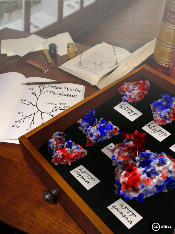
About the image: Similar to early comparisons of different species that led to the establishment of phylogenetic trees, high resolution structural information can be used to identify unique as well as shared structural properties that can be explored and cluster members of a enzyme family into a tree based on molecular properties. The structural comparison of PTPs revealed that many features cannot be reliably predicted from sequence analysis alone. The phosphatase images are coloured according to the surface electrostatic potential using ICM.
The human genome contains 107 PTPs grouped into four distinct families: Class I cysteine-based PTPs constitute the largest family. They are divided into 38 “classical” tyrosine specific PTPs and 61 dual specificity phosphatases (DUSPs) [4]. The classical PTPs are one of the most comprehensively structurally covered protein families: 32 structures of the 49 D1/D2 domains are currently available. PTPs share a highly conserved overall fold but have very diverse protein surface properties.
4. Alonso et al.. Protein tyrosine phosphatases in the human genome. Cell (2004) 117(6):699-711.
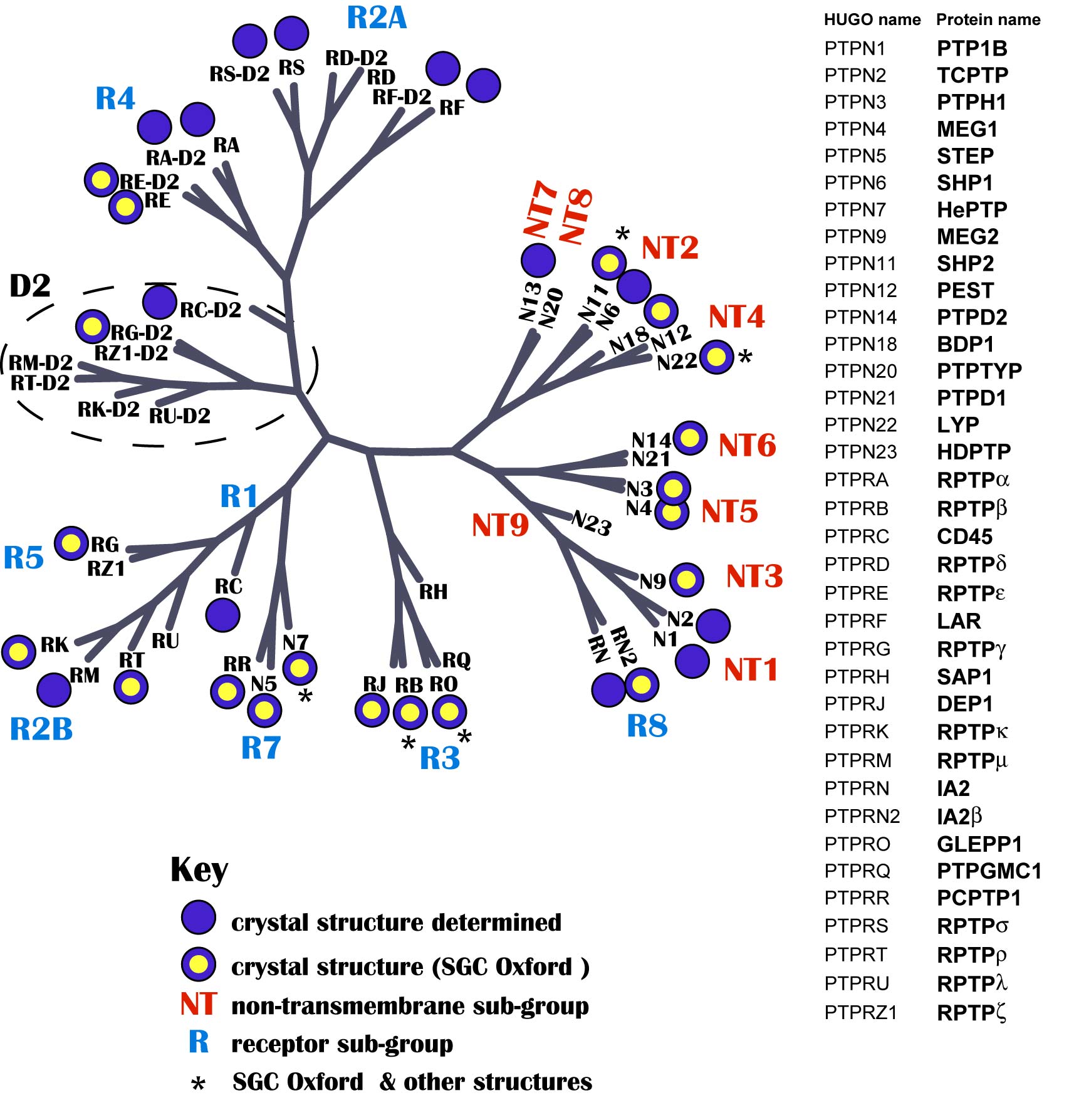
Structural coverage of the human PTP family: Catalytic domains with known three dimensional structure are highlighted by a dot. Dots with a yellow center indicate structures released by our laboratory.
PDB code | Release date | Phosphatase | Species | Description | Sector |
|---|---|---|---|---|---|
| 3EU0 | 11.Nov.2008 | PTPN1 | Human | Crystal structure of the S-nitrosylated Cys215 of PTP1B | Academia |
| 2ZMM | 07.Oct.2008 | PTPN1 | Human | Crystal structure of PTP1B-inhibitor complex, 4-bromo-3-(carboxymethoxy)- 5-{3-[cyclohexyl (methylcarbamoyl) amino]phenyl} thiophene-2- carboxylic acid | Industry |
| 2ZN7 | 07.Oct.2008 | PTPN1 | Human | Crystal structure of PTP1B-inhibitor complex, 4-bromo-3-(carboxymethoxy)- 5-{3-[cyclohexyl (phenylcarbonyl) amino]phenyl} thiophene-2- carboxylic acid | Industry |
| 3D9C | 23.Sep.2008 | PTPN1 | Human | Crystal Structure PTP1B complex with aryl Seleninic acid | Academia |
| 2QDC | 24.Jun.2008 | PTPN7 | Human | Crystal structure of the HePTP catalytic domain D236A mutant | Academia |
| 2QDM | 24.Jun.2008 | PTPN7 | Human | Crystal structure of the HePTP catalytic domain C270S/D236A/Q314A mutant | Academia |
| 2QDP | 24.Jun.2008 | PTPN7 | Human | Crystal structure of the HePTP catalytic domain C270S mutant crystallized in ammonium acetate | Academia |
| 3CWE | 10.Jun.2008 | PTPN1 | Human | PTP1B in complex with a phosphonic acid inhibitor | Industry |
| 2JJD | 08.Apr.2008 | PTPRE | Human | pProtein tyrosine phosphatase, receptor type, E isoform | SGC Oxford |
Click on the protein name to read more about the phosphatase and its structure in our website. To explore and view the structure in your web browser, please click on the "iSee" icon (first-time user: you'll be asked to install our visualisation plug-in - please follow the installation instructions that will be displayed).
HUGO name | Protein Name | PDB code | ||
 | RPTPκ | 2C7S | ||
 | RPTPρ | 2OOQ | ||
 | RPTPε | 2JJD | coming soon | |
 | RPTPγ | 2NLK | ||
 | RPTPγ | 2H4V | ||
 | RPTPβ | 2AHS | ||
 | DEP1 | 2CFV, 2NZ6 | ||
 | GLEPP1 | 2GJT | ||
 | PCPTP1 | 2A8B |
| |
 | STEP | 2BIJ | ||
 | STEP | 2CJZ | ||
| STEP | 2BV5 |
HUGO name | Protein Name | PDB code | ||
 | HePTP | 2A3K | ||
 | IA2β | 2QEP | ||
 | SHP2 | 3B7O | ||
 | MEG2 | 2PA5 | ||
 | PTPH1 | 2B49 | ||
 | MEG1 | 2I75 | ||
 | BDP1 | 2OC3 | ||
 | LYP | 2P6X | ||
 | PTPD2 | 2BZL |