Skip to main content
About
About SGC
Members & Technology Partners
Governance
Open Science
Impact
SGC Gender Equality Plan
SGC Sites & People
Global
SGC Toronto
SGC UNC
SGC Karolinska
SGC Frankfurt
SGC Neuro
SGC UCL
SGC Unicamp
Science and Resources
Proteins
Structure Gallery
Protein Production Resources
Expression Vectors
Constructs
Legacy Protein Protocols
Chemistry
Chemical Probes
SGC Chemical Probes
Donated Chemical Probes
Chemical Handles
Chemogenomic Sets
Open Data
Target Enabling Packages
Patient Tissue Platform
Open Lab Notebooks
Publications
Bioinformatics Resources
ChemBioPort
UbiHub
ChromoHub
Initiatives
Overview
Target 2035
Protein Contribution Network
AIRCHECK
MAINFRAME
CACHE
Conscience
Agora Open Science Trust
EUbOPEN
Open Chemistry Networks
Women’s and Children’s Health Initiative
READDI AViDD
TREAT AD
YCharOS
News & Outreach
News from SGC
Press Releases
Blogs
Get Involved
Participate
Donate
Careers
Contact Us
Donate
Home
About
About SGC
Members & Technology Partners
Governance
Open Science
Impact
SGC Gender Equality Plan
SGC Sites & People
Global
SGC Toronto
SGC UNC
SGC Karolinska
SGC Frankfurt
SGC Neuro
SGC UCL
SGC Unicamp
Science and Resources
Proteins
Structure Gallery
Protein Production Resources
Expression Vectors
Constructs
Legacy Protein Protocols
Chemistry
Chemical Probes
SGC Chemical Probes
Donated Chemical Probes
Chemical Handles
Chemogenomic Sets
Open Data
Target Enabling Packages
Patient Tissue Platform
Open Lab Notebooks
Publications
Bioinformatics Resources
ChemBioPort
UbiHub
ChromoHub
Initiatives
Overview
Target 2035
Protein Contribution Network
AIRCHECK
MAINFRAME
CACHE
Conscience
Agora Open Science Trust
EUbOPEN
Open Chemistry Networks
Women’s and Children’s Health Initiative
READDI AViDD
TREAT AD
YCharOS
News & Outreach
News from SGC
Press Releases
Blogs
Get Involved
Participate
Donate
Careers
Contact Us
Donate
Search
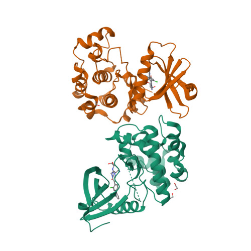
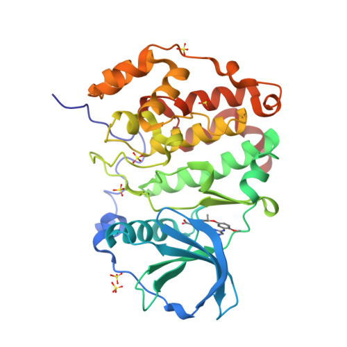
Protein:
STK24
PDB ID:
8QLR
Deposition Date:
Wednesday 20th September 2023
Authors:
Balourdas, D.I., Rak, M., Knapp, S., Joerger, A.C.
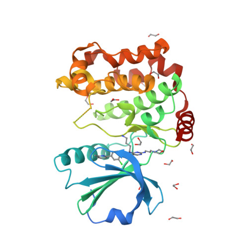
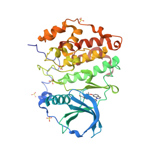
Protein:
STK24
PDB ID:
8QLS
Deposition Date:
Wednesday 20th September 2023
Authors:
Balourdas, D.I., Rak, M., Knapp, S., Joerger, A.C.
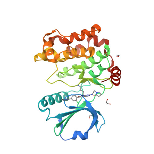
Protein:
STK24
PDB ID:
8QLT
Deposition Date:
Wednesday 20th September 2023
Authors:
Balourdas, D.I., Rak, M., Knapp, S., Joerger, A.C.
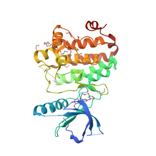
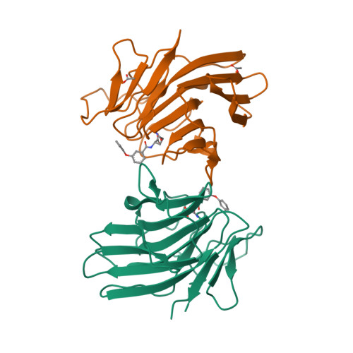
Protein:
EPHA2
PDB ID:
8QQY
Deposition Date:
Friday 6th October 2023
Authors:
Zhubi, R., Gerninghaus, J., Knapp, S., Kraemer, A.
Protein:
CSK2a1
PDB ID:
8QWY
Deposition Date:
Friday 20th October 2023
Authors:
Kraemer, A., Galal, K., Willson, T., Knapp, S.
Protein:
CSK2a1
PDB ID:
8QWZ
Deposition Date:
Friday 20th October 2023
Authors:
Kraemer, A., Galal, K., Willson, T., Knapp, S.
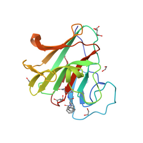
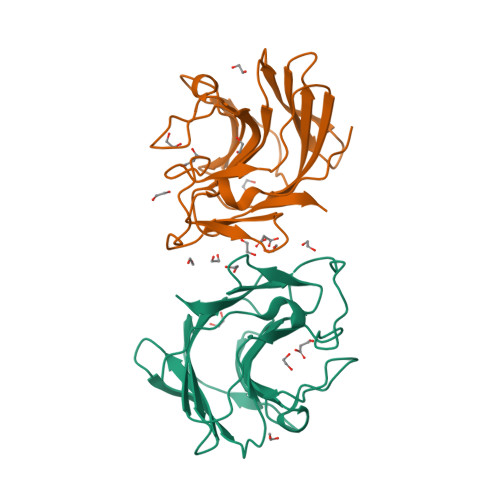
Protein:
TRIM7
PDB ID:
8R5B
Deposition Date:
Thursday 16th November 2023
Authors:
Munoz Sosa, C.J., Kraemer, A., Knapp, S.
Protein:
TRIM7
PDB ID:
8R5C
Deposition Date:
Thursday 16th November 2023
Authors:
Munoz Sosa, C.J., Kraemer, A., Knapp, S.
Protein:
TRIM7
PDB ID:
8R5D
Deposition Date:
Thursday 16th November 2023
Authors:
Munoz Sosa, C.J., Kraemer, A., Knapp, S.
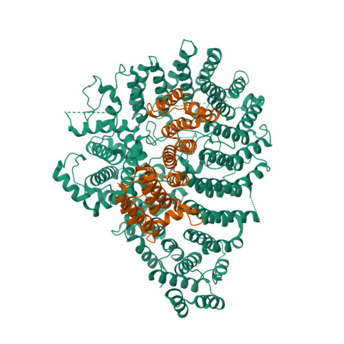
Protein:
HTT
PDB ID:
8SAH
Deposition Date:
Friday 31st March 2023
Authors:
Harding, R.J., Deme, J.C., Alteen, M.G., Arrowsmith, C.H., Lea, S.M.
Pagination
First page
« First
Previous page
‹ Previous
…
Page
458
Page
459
Page
460
Page
461
Page
462
Page
463
Page
464
Page
465
Page
466
…
Next page
Next ›
Last page
Last »
Subscribe to