VinSpinIn A chemical probe for the Spin family proteins

overview
Probe Negative control
 

VinSpinIn (Vinnie's Spindlin Inhibitor)


VinSpinIC (Vinnie's Spindlin Inactive Control)

The Spindlin proteins are tudor domain containing proteins. There are five spindlin family members (Spin1, Spin2A, Spin2B, Spin3, Spin4), which are expressed at various levels throughout the body. However, spindlin1 (Spin1) is expressed at higher levels compared to the other family members.1 Our current knowledge of the biological roles of spindlins is limited to that of Spin1.

The Spin proteins consist of three tudor-like Spin/Ssty domains, arranged in a clockwise orientation.2-4 Spin1 has been shown to bind to trimethylated lysine 4 of histone 3 (H3K4me3) via domain 2 (ITC Kd = 147 nM).3,5 However, increased binding is observed when a second epigenetic methylation mark is present on arginine 8 (asymmetrically dimethylated) (H3K4me3R8me2a; ITC Kd = 45 nM).2 Spin1 also binds to H4K20me3 via domain 2.6

Spin1 binding to methylated histones is associated with transcriptional activation. Spin1 was found to be overexpressed in various cancers and has been shown to drive cancer cell proliferation through activation of the Wnt/β-catenin, PI3K/Akt and RET signalling pathways.2,7-10

Conversely, Spin1 has been shown to facilitate the inactivation of p53 by sequestering the ribosomal protein uL18.11

Direct or indirect Spin1 knockdown resulted in cancer cell and xenograft tumor growth inhibition, and such studies suggest that small molecule inhibition of Spin1 may be a viable approach for the treatment of certain cancers.8-13

Therefore, the SGC has developed VinSpinIn as a potent, cell active chemical probe for the Spin family proteins. VinSpinIn, along with the structurally very similar inactive control compound VinSpinIC, will contribute significantly to the elucidation of the biological roles and functions of the spindlin proteins, and will aid with the validation of Spin1 as a chemotherapeutic target.

Potency Against Target Family
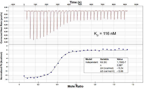
The SYPRO Orange thermal shift assay was employed to assess the potency of VinSpinIn and VinSpinIC against four of the five Spin family members (Table 1). VinSpinIn induced a large shift in the thermal stability in all of the Spin protein assessed, while VinSpinIC did not. ITC was also performed to determine the potency of VinSpinIn on four Spin family members as well as an additional Spin1 construct (Table 1). VinSpinIn had KDs ranging between approximately 10-130 nM across the family.

Spin Family Proteins

Thermal Shift Assay

(ΔTm°C)

ITC Assay

(KD nM)

VinSpinInVinSpinICVinSpinIn
Spin149-262Not testedNot tested9.9
SPIN126-26213.171.02111.1
SPIN2B22-25810.470.2946.1
SPIN321-25814.122.34131.1
SPIN436-2496.530.2518.1

Table 1: Potency Against Target Family

Selectivity
VinSpinIn and VinSpinIC were screened against a panel of methyl binding domains (MBDs) using the thermal shift assay (Table 2). No significant thermal shift was observed for any of the MBDs screened.
A Scintillation Proximity Assay (SPA) was employed to screen VinSpinIn and VinSpinIC against a panel of methyltransferases and IC50s were determine for selected targets (Table 3). The lowest IC50 of VinSpinIn (PRMT4) was approximately 300 times greater than the AlphaScreen IC50 on Spin1 (30 nM).

Methyl Lysine Binders

VinSpinIn

(ΔTm°C)

VinSpinIC

(ΔTm°C)

UHRF1139-298-0.04-0.14
53BP11483-1606-0.65-0.41
TDRD3525-6110.15-0.95
SND1650-910-0.09-1.62
SETDB1197-403-0.04-0.34
SGF29129-293-0.05-0.92
CCDC101114-293-0.040.3
CHD1269-446-0.652.37
FALZ2736-27930.150.31
ING2211-266-0.090.25
JARID1A1542-1660-0.04-0.04
MLL1558-1773-0.050.63
MLL5113-171-0.05-0.05
PHF21-640.020.19
PHF81-630.23-0.11
TAF31086-1153-0.21-0.04

Table 2: Screening on a panel of MBDs.

Methyltransferase Panel

VinSpinIn

(IC50 µM)

VinSpinIC

(IC50 µM)

PRMT492
SETD2205
PRMT7198
SUV39H13418
PRMT62719
PRC22127
SMYD22244
PRDM92544
PRMT14759
PRMT84566

Table 3: IC50s on methyltransferases.

Dosage
Use between 0.5 and 3 µM for Cellular Assays and 1 µM for screening at a single shot, for both VinSpinIn & VinSpinIC.

In vitro Activity
In a number of biophysical assays, VinSpinIn was shown to be a potent Spin1 inhibitor and has an ITC KD which is approximately 130 times more potent than that of the inactive control VinSpinIC (Table 4).

Compound

AlphaScreen

(Spin126-262)

(IC50)

Octet BLI

(Spin126-262)

(KD)

ITC

(Spin126-262)

(KD)

ITC

(Spin149-262)

(KD)

Tm Shift

(Spin126-262)

(ΔTm°C)

VinSpinIn30 nM55 nM111.1 nM9.9 nM13.2
VinSpinIC3.64 µMnot testednot tested1.3 µM1.0

Table 4: In vitro biophysical binding assay results of VinSpinIn & VinSpinIC.

Cellular Activity
In a NanoBRET cellular target engagement assay VinSpinIn displayed dose dependant inhibition of the Spin1-H3 interaction; the inactive VinSpinIC showed no inhibition (Figure 2).


Figure 2: NanoBRET cellular engagement assay. Click on the ‘Cell-based Assay Data’ tab above for more details.

properties
VinSpinIn

Click here to download the SDF file.

VinSpinIn
Physical and chemical properties
Molecular weight738.96 g/mol
Molecular formulaC42H58N8O4
IUPAC name2-[4-(2-{[2-(3-{[2-amino-5-(cyclopropylmethoxy)-3,3-dimethyl-3H-indol-6-yl]oxy}propyl)-2,3-dihydro-1H-isoindol-5-yl]oxy}ethyl)-1H-1,2,3-triazol-1-yl]-1-{4-[2-(pyrrolidin-1-yl)ethyl]piperidin-1-yl}ethan-1-one
logP3.85
TPSA123.57
No. of chiral centres0
No. of rotatable bonds17
No. of hydrogen bond acceptors2
No. of hydrogen bond donors12
Storage+4 °C
DissolutionDMSO (up to at least 50 mM)
VinSpinIC

Click here to download the SDF file.

VinSpinIC
Physical and chemical properties
Molecular weight738.96 g/mol
Molecular formulaC42H58N8O4
IUPAC name2-[4-({[2-(4-{[2-amino-5-(cyclopropylmethoxy)-3,3-dimethyl-3H-indol-6-yl]oxy}butyl)-2,3-dihydro-1H-isoindol-5-yl]oxy}methyl)-1H-1,2,3-triazol-1-yl]-1-{4-[2-(pyrrolidin-1-yl)ethyl]piperidin-1-yl}ethan-1-one
clogP3.99
TPSA123.57
No. of chiral centres0
No. of rotatable bonds17
No. of hydrogen bond acceptors2
No. of hydrogen bond donors12
Storage+4 °C
DissolutionDMSO (up to at least 50 mM)

SMILES:
VinSpinIn: CC1(C(N)=NC2=CC(OCCCN3CC4=C(C3)C=C(OCCC5=CN(N=N5)CC(N6CCC(CC6)CCN7CCCC7)=O)C=C4)=C(C=C21)OCC8CC8)C
VinSpinIC:  O=C(CN1N=NC(COC2=CC3=C(C=C2)CN(C3)CCCCOC4=CC5=C(C(C)(C(N)=N5)C)C=C4OCC6CC6)=C1)N7CCC(CC7)CCN8CCCC8

InChI:
VinSpinIn: InChI=1/C42H58N8O4/c1-42(2)36-23-38(54-29-31-6-7-31)39(24-37(36)44-41(42)43)53-20-5-16-48-25-32-8-9-35(22-33(32)26-48)52-21-13-34-27-50(46-45-34)28-40(51)49-18-11-30(12-19-49)10-17-47-14-3-4-15-47/h8-9,22-24,27,30-31H,3-7,10-21,25-26,28-29H2,1-2H3,(H2,43,44)/f/h43H2
VinSpinIC: InChI=1/C42H58N8O4/c1-42(2)36-22-38(54-28-31-7-8-31)39(23-37(36)44-41(42)43)52-20-6-5-16-48-24-32-9-10-35(21-33(32)25-48)53-29-34-26-50(46-45-34)27-40(51)49-18-12-30(13-19-49)11-17-47-14-3-4-15-47/h9-10,21-23,26,30-31H,3-8,11-20,24-25,27-29H2,1-2H3,(H2,43,44)/f/h43H2

InChIKey:
VinSpinIn: XPEJZXWPKDAYFX-UHFFFAOYSA-N
VinSpinIC: FOBGCBSESHUBEI-UHFFFAOYSA-N

Supplemental Material

Experimental_VinSpinIn, VinSpinIn Spectra

selectivity profile

The SYPRO Orange thermal shift assay was employed to screen VinSpinIn and VinSpinIC against a panel of methyl binding domains (MBDs), which included four of the five Spin family members (Table 1). VinSpinIn induced a large shift in thermal stability of Spin1, Spin2B, Spin3, Spin4. No other significant shift in thermal stability was observed for VinSpinIn or the inactive VinSpinIC.

Methyl Lysine Binders

VinSpinIn

(ΔTm°C)

VinSpinIC

(ΔTm°C)

a UHRF1139-298-0.04-0.14
a 53BP11483-1606-0.65-0.41
a TDRD3525-6110.15-0.95
a SND1650-910-0.09-1.62
a SETDB1197-403-0.04-0.34
a SGF29129-293-0.05-0.92
b CCDC101114-293-0.040.3
b CHD1269-446-0.652.37
b FALZ2736-27930.150.31
b ING2211-266-0.090.25
b JARID1A1542-1660-0.04-0.04
b MLL1558-1773-0.050.63
b MLL5113-171-0.05-0.05
b PHF21-640.020.19
b PHF81-630.23-0.11
b TAF31086-1153-0.21-0.04
b SPIN126-26213.171.02
b SPIN2B22-25810.470.29
b,c SPIN321-25814.122.34
b SPIN436-2496.530.25

Table 1: Screening on a panel of MBDs.a protein = 0.05 to 0.2 mg/ml, compound = 200 µM; b protein = 2 µM, compound = 20 µM; c Intrinsic tryptophan fluorescence used

A fluorescence polarization displacement assays was employed to screen the compounds against L3MBTL1 and L3MBTL3 (MBT methyl lysine readers), which gave respective Kdisp.s of 27 and 8 µM for VinSpinIn, and 28 and 14 µM for the inactive VinSpinIC. Therefore, VinSpinIn is approximately 267 times less potent towards L3MBTL1 than Spin1.

Selectivity screening was also performed against a panel of methyl transferases domains (MTDs) including protein, DNA and RNA methyltransferases using a Scintillation Proximity Assay (SPA) (Figure 1).

Figure 1: (Top) Methyltransferase activity in presence of VinSpinIn at 10 & 50 µM
(Bottom) Methyltransferase activity in presence of VinSpinIC at 10 and 50 µM.

For selected targets IC50s were determined (Table 2). The lowest IC50 of VinSpinIn (PRMT4) was approximately 300 times greater than the AlphaScreen IC50 on Spin1.

Methyltransferase Panel

VinSpinIn

(IC50 µM)

VinSpinIC

(IC50 µM)

PRMT492
SETD2205
PRMT7198
SUV39H13418
PRMT62719
PRC22127
SMYD22244
PRDM92544
PRMT14759
PRMT84566
DNMT3A/3La9
DNMT3B/3La5
G9aa9
GLPa21
SETDB1a6

Table 2: IC50s of selected methyltransferases.a Not determined

Materials and Methods

Effects of VinSpinIn  and VinSpinIC on methyltransferase activity of G9a, GLP, SUV39H1, SUV39H2, SETDB1, SETD8, SUV420H1, SUV420H2, SETD7, MLL1 trimeric complex, MLL3 pentameric complex, EZH2 trimeric complex, PRMT1, PRMT3, PRMT4, PRMT5-MEP50 complex, PRMT6, PRMT7, PRMT8, PRMT9, PRDM9, SETD2, SMYD2, SMYD3, and DNMT1 was assessed by monitoring the incorporation of tritium-labeled methyl group to lysine or arginine residues of peptide substrates using Scintillation Proximity Assay (SPA) as previously described14.  Assays were performed in a 10 µl reaction mixture containing 3H-SAM (Cat.# NET155V250UC; Perkin Elmer; www.perkinelmer.com) at substrate concentrations close to Km values for each enzyme.  Two concentrations (10µM and 50 µM) of VinSpinIn or  VinSpinIC  were used in all selectivity assays.  To stop the enzymatic reactions, 10 µl of 7.5 M guanidine hydrochloride was added, followed by 180 µl of buffer (20 mM Tris, pH 8.0), mixed and then transferred to a FlashPlate (Cat.# SMP103; Perkin Elmer; www.perkinelmer.com). After mixing, the reaction mixtures in Flash plates were incubated for 2 hours and the CPM were measured using Topcount plate reader (Perkin Elmer, www.perkinelmer.com). The CPM counts in the absence of compound for each data set were defined as 100% activity. In the absence of the enzyme, the CPM counts in each data set were defined as background (0%).

For DOT1L, NSD1, NSD2, NSD3, ASH1L, DNMT3A/3L, and DNMT3B/3L, a filter-based assay was used. In this assay, 10 µl of reaction mixtures were incubated at 23 oC for 1 hour, 50 µl of 10% trichloroacetic acid (TCA) was added, mixed and transferred to filter-plates (Millipore; cat.# MSFBN6B10; www.millipore.com). Plates were centrifuged at 2000 rpm (Allegra X-15R - Beckman Coulter, Inc.) for 2 min followed by 2 additional 10% TCA wash and one ethanol wash followed by centrifugation. Plates were dried and 30 µl MicroO (MicroScint-O; Cat.# 6013611, Perkin Elmer; www.perkinelmer.com) was added to each well, centrifuged and removed. 50 µl of MicroO was added again and CPM was measured using Topcount plate reader.

IC50 Determinations:
IC50 values were determined for inhibition of methyltransferase activity of the following enzymes:

  • PRMT4 (20 nM PRMT4, 1 µM of biotinlated-H3 1-25, 2 µM of 3H-SAM)
  • SETD2 (150 nM SETD2, 0.5 µM of biotinylated-H3 21-44, 5 µM of 3H-SAM)
  • PRMT7 (25 nM PRMT7, 0.3 µM of biotinylated-H2B(23-37), 1 µM of 3H-SAM)
  • SUV39H1 (20 nM SUV30H1, 0.2 µM of biotinlated-H3 1-25, 5 µM of 3H-SAM)
  • PRMT6 (20 nM PRMT6, 1 µM of biotinlated-H4 1-24, 2 µM of 3H-SAM)
  • PRC2 (20 nM PRC2, 1 µM of biotinlated-H3 21-44, 2 µM of 3H-SAM)
  • SMYD2 (30 nM SMYD2, 3 µM of biotinlated-P53peptide, 0.3 µM of 3H-SAM)
  • PRDM9 (5 nM PRDM9, 4 µM of biotinlated-H3 1-25, 71 µM of SAM)
  • PRMT1 (15 nM PRMT1, 0.13 µM of biotinlated-H4 1-24, 5 µM of 3H-SAM)
  • PRMT8 (20 nM PRMT8, 1 µM of biotinlated-H4 1-24, 2 µM of 3H-SAM)
  • DNMT3A/3L (20 nM DNMT3A/3L, 0.5 µM of poly DI-DC, 1 µM of 3H-SAM)
  • DNMT3B/3L (50 nM DNMT3B/3L, 0.5 µM of poly DI-DC, 1 µM of 3H-SAM)
  • G9a (5 nM G9A, 0.8 µM of biotinlated-H3 1-25, 10 µM of SAM)
  • GLP (5 nM Glp, 0.8 µM of biotinlated-H3 1-25, 10 µM of SAM).

To stop the enzymatic reactions, 7.5 M Guanidine hydrochloride was added, followed by 180 µL of buffer (20 mM Tris, pH 8.0), mixed and then transferred to a FlashPlate (Cat.# SMP103; Perkin Elmer; www.perkinelmer.com). After mixing, the reaction mixtures in Flash plate were incubated for 2 hour and the CPM counts were measured using Topcount plate reader (Perkin Elmer, www.perkinelmer.com). The CPM counts in the absence of compound for each data set were defined as 100% activity. In the absence of the enzyme, the CPM counts in each data set were defined as background (0%). The IC50 values were calculated using GraphPad Prism 7 software.

in vitro potency
cell based assay data

In a NanoBRET cellular target engagement assay VinSpinIn displayed dose dependant inhibition of the Spin1-H3 interaction, with an IC50 of 270 nM. The inactive VinSpinIC showed no inhibition (Figure 1).

Figure 1: NanoBRET cellular target engagement assay performed in U2OS cells. Full length SPIN1 with N-terminal NanoLuc; Histone 3.3 with C-terminal Halotag; 24h incubation with VinSpinIn and VinSpinIC

Materials and Methods

U20S cell (2.8 x 10⁵) were plated in each well of a 6-well plate after 6h cells were co-transfected with C-terminal HaloTag-Histone 3.3 (NM_002107) and an N-terminal NanoLuciferase fusion of full length SPIN1 at a 1:500 (NanoLuc® to HaloTag®) ratio respectively with FuGENE HD transfection regent15.

Sixteen hours post-transfection, cells were collected, washed with PBS, and exchanged into media containing phenol red-free DMEM and 4% FBS in the absence (control sample) or the presence (experimental sample) of 100 nM NanoBRET 618 fluorescent ligand (Promega). Cells were then re-plated in a 384-well assay white plate (Greiner #3570) at 2.7x10³ cells per well.  VinSpinIn and VinSpinIC were then added directly to media at final concentrations 0-30μM or an equivalent amount of DMSO as a vehicle control, and the plates were incubated for 24 h at 37oC in the presence of 5% CO2.

NanoBRET Nano-Glo substrate (Promega) was added to both control and experimental samples at a final concentration of 10 µM.  Readings were performed within 10 minutes using a ClarioSTAR (BMG labtech) equipped with 460 nm and 610 nm filters.  A corrected BRET ratio was calculated and is defined as the ratio of the emission at 610 nm/460 nm for experimental samples minus the emission at 610 nm/460 nm for control samples (without NanoBRET fluorescent ligand). BRET ratios are expressed as milliBRET units (mBU), where 1 mBU corresponds to the corrected BRET ratio multiplied by 1000.

references
  1. https://www.proteinatlas.org/search/Spindlin.
  2. Su, X. et al. Molecular basis underlying histone H3 lysine-arginine methylation pattern readout by Spin/Ssty repeats of Spindlin1. Genes Dev 28, 622-636, doi: 10.1101/gad.233239.113 (2014).
  3. Yang, N. et al. Distinct mode of methylated lysine-4 of histone H3 recognition by tandem tudor-like domains of Spindlin1. Proc Natl Acad Sci U S A 109, 17954-17959, doi: 10.1073/pnas.1208517109 (2012).
  4. Zhao, Q. et al. Structure of human spindlin1. Tandem tudor-like domains for cell cycle regulation. J Biol Chem 282, 647-656, doi: 10.1074/jbc.M604029200 (2007).
  5. Wang, W. et al. Nucleolar protein Spindlin1 recognizes H3K4 methylation and stimulates the expression of rRNA genes. EMBO Rep 12, 1160-1166, doi: 10.1038/embor.2011.184 (2011).
  6. Shanle, E. K. et al. Histone peptide microarray screen of chromo and Tudor domains defines new histone lysine methylation interactions. Epigenetics Chromatin 10, 12, doi: 10.1186/s13072-017-0117-5 (2017).
  7. Wang, J. X. et al. SPINDLIN1 Promotes Cancer Cell Proliferation through Activation of WNT/TCF-4 Signaling. Mol Cancer Res 10, 326-335, doi: 10.1158/1541-7786.Mcr-11-0440 (2012).
  8. Franz, H. et al. The histone code reader SPIN1 controls RET signaling in liposarcoma. Oncotarget 6, 4773-4789, doi: 10.18632/oncotarget.3000 (2015).
  9. Chen, X. et al. Suppression of SPIN1-mediated PI3K-Akt pathway by miR-489 increases chemosensitivity in breast cancer. J Pathol 239, 459-472, doi: 10.1002/path.4743 (2016).
  10. Li, Y., Ma, X., Wang, Y. & Li, G. miR-489 inhibits proliferation, cell cycle progression and induces apoptosis of glioma cells via targeting SPIN1-mediated PI3K/AKT pathway. Biomed Pharmacother 93, 435-443, doi: 10.1016/j.biopha.2017.06.058 (2017).
  11. Fang, Z. et al. SPIN1 promotes tumorigenesis by blocking the uL18 (universal large ribosomal subunit protein 18)-MDM2-p53 pathway in human cancer. Elife 7, doi: 10.7554/eLife.31275 (2018).
  12. Chen, X. et al. Long noncoding RNA MHENCR promotes melanoma progression via regulating miR-425/489-mediated PI3K-Akt pathway. Am J Transl Res 9, 90-102 doi: 10.1042/BSR20170682 (2017).
  13. Drago-Ferrante, R. et al. Suppressive role exerted by microRNA-29b-1-5p in triple negative breast cancer through SPIN1 regulation. Oncotarget 8, 28939-28958, doi: 10.18632/oncotarget.15960 (2017).
  14. Barsyte-Lovejoy, D. et al. (R)-PFI-2 is a potent and selective inhibitor of SETD7 methyltransferase activity in cells, PNAS 111, 12853-12858. doi: 10.1073/pnas.1407358111 (2014).
  15. Machleidt, T. et al. NanoBRET--A Novel BRET Platform for the Analysis of Protein-Protein Interactions. ACS Chem Biol 10, 1797-1804, doi: 10.1021/acschembio.5b00143 (2015).
pk properties
co-crystal structures

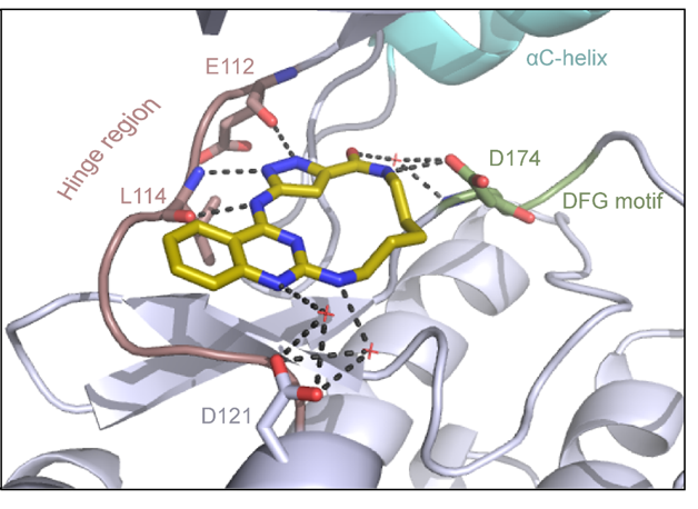
The co-crystal structure of Spin1 with VinSpinIn confirms that VinSpinIn binds to both domain 1 and 2 of Spin1, and therefore is referred to as a bidentate inhibitor (Figure 1).

Figure 1: Co-crystal structure of Spin1 (domain 1 = purple; domain 2 = green; domain 3 = yellow) with VinSpinIn (Cyan).

synthetic schemes
materials and methods

DDR-TRK-1 A chemical probe for the DDR and TRK kinase

The probe DDR-TRK-1 is available from Cayman Chemical and Sigma.

overview
Probe Negative control


DDR-TRK-1


DDR-TRK-1N

The discoidin domain receptors (DDRs), DDR1 and DDR2, are unique among the receptor tyrosine kinases (RTKs) in being activated by interaction with the extracellular matrix via binding to triple-helical collagen by the receptor extracellular domains.1  DDR1 and DDR2 form constitutive dimers making them unusual among RTKs, which typically dimerize only upon activation.2 DDRs regulate extracellular matrix remodeling, as well as cell adhesion, proliferation and migration.3 DDR kinases are linked to the progression of various human diseases, including fibrotic disorders, atherosclerosis and cancer.3-5  Significantly, they are identified as indicators of poor prognosis in ovarian, breast and lung cancer.6 DDR1 overexpression is associated with increased cell survival and invasion in hepatocellular carcinomas, pituitary adenoma and prostate cancer,7 whereas DDR2 is mutated in squamous cell lung cancers8 and contributes to breast cancer metastasis.9 The promise of DDR kinases as a therapeutic target has been demonstrated by DDR1 knockdown that has been shown to reduce metastatic activity in lung cancer models,10 slow the development of atherosclerosis,5 and impede the development of fibrotic disorders.11

The TRK kinases are represented by three members, TRKA, TRKB, and TRKC, which are selectively expressed in neuronal tissue.  Receptor signaling is initiated by binding of the neurotrophic factors NGF, BDNF, and NTR, respectively.  Subsequent signaling is via the RAS, PLCγ and PI3γ pathways.  The TRKs play a vital role in CNS development and survival. Gene fusions, protein overexpression, and single nucleotide alterations, have been implicated in the pathogenesis of specific cancer types including glioblastoma, papillary thyroid carcinoma, and secretory breast carcinomas, but are rare in most other cancers.12

DDR-TRK-1 is a chemical probe for the DDR and TRK kinases with good in vitro and in vivo potency.
DDR-TRK-1 inhibits colony formation and migration of Panc-1 pancreatic cancer cells.  In cellular and mouse models of lung fibrosis, DDR-TRK-1 inhibits signaling, expression of fibrotic markers and fibrotic features such as hydroxproline expression.  With restricted CNS exposure and therefore no TRK inhibition, the in vivo effects of DDR-TRK-1 can be attributed to DDR1-2 inhibition. DDR-TRK-1N is the negative control compound with very minimal differences in compound structure, and should be used in parallel to DDR-TRK-1. The probe can be complemented by the use of BAY-826, which inhibits the kinases TIE1, TIE2, DDR1 and DDR2, but lacks TRK activity to better understand the target involved in the phenotypic effect.

Work on this probe has been published in J.Med.Chem. 2016 59(12), p 5911-5916, 'Structure-Based Design of Tetrahydroisoquinoline-7-carboxamides as Selective Discoidin Domain Receptor 1 (DDR1) inhibitors'.

Potency Against Target Family

DDR-TRK-1 is a chemical probe for the DDR and TRK kinases (IC50 3-43 nM) with corresponding good cellular potency in NanoBRETTM target engagement assays (IC50 104-448 nM).  DDR-TRK-1 was shown to be selective in an in vitro kinase panel followed by cellular NanoBRETTM assays.

Selectivity

DDR-TRK-1 is selective in KINOMEscan® at 1μM. The closest off target is CDK11 (370 nM), which was however shown to be only poorly inhibited in cells  (~5 µM in NanoBRETTM). The negative control DDR-TRK-1N is entirely clean in KINOMEscan at 1μM.

Dosage

We recommend that DDR-TRK-1 be used at 5μM concentration in cells. The negative control DDR-TRK-1N should be used at 5μM concentration in cells – it is toxic in HeLa cells above 10μM.

We also recommend the use of selective TRK inhibitors or the TIE-DDR BAY-826 probe in parallel to dissect the biology of DDR1/2 versus TRKA/B/C.

Cellular Activity

In NanoBRETTM assays, DDR-TRK-1 shows a potency of 104nM against DDR1, 175nM against DDR2, 448nM against TRKA and 142nM against TRKB.

In vitro Activity

In an activity assay at RBC (10μM ATP), DDR-TRK-1 shows an IC50 value of 27nM against DDR1, 4.5nM against DDR2, 43nM against TRKA, 3.6nM against TRKB and 2.9nM against TRKC.

properties
Probe Negative control


DDR-TRK-1


DDR-TRK-1N

Click here to download the DDR-TRK-1 SDF file. Click here to download the DDR-TRK-1N SDF file.
Physical and chemical properties for DDR-TRK1
Molecular weight492.1885
Molecular formulaC29 H31 F3 N4 O
IUPAC name(3-(4-methyl-1H-imidazol-1-yl)-5-(trifluoro-methyl)-phenylamino)-(5-methyl-3-(pyrimidin-5-yl)-3-aza-bicyclo[4.4.0]deca-1(6),7,9-trien-9-yl)-methanone
MollogP4.6
PSA59.2
No. of chiral centres1
No. of rotatable bonds6
No. of hydrogen bond acceptors5
No. of hydrogen bond donors1
Storage-20 as DMSO stock
DissolutionSoluble in DMSO at least up to 50mM
Physical and chemical properties for DDR-TRK-1N
Molecular weight508.2450
Molecular formulaC26 H23 F3 N6 O
IUPAC name(3-((4-methyl-piperazin-1-yl)-methyl)-5-(trifluoro-methyl)-phenylamino)-(3-phenyl-3-aza-bicyclo[4.4.0]deca-1(6),7,9-trien-9-yl)-methanone
MollogP5.6
PSA33.6531
No. of chiral centres0
No. of rotatable bonds7
No. of hydrogen bond acceptors4
No. of hydrogen bond donors1
Storage-20 as DMSO stock
DissolutionSoluble in DMSO at least up to 50mM

SMILES:
DDR-TRK-1: CC1=CN(C2=CC(NC(C3=CC=C4C(CN(C5=CN=CN=C5)C[C@@H]4C)=C3)=O)=CC(C(F)(F)F)=C2)C=N1
DDR-TRK-1N: CN1CCN(CC2=CC(C(F)(F)F)=CC(NC(C3=CC4=C(CCN(C5=CC=CC=C5)C4)C=C3)=O)=C2)CC1

InChI:
DDR-TRK-1: InChI=1S/C26H23F3N6O/c1-16-11-34(23-9-30-14-31-10-23)13-19-5-18(3-4-24(16)19)25(36)33-21-6-20(26(27,28)29)7-22(8-21)35-12-17(2)32-15-35/h3-10,12,14-16H,11,13H2,1-2H3,(H,33,36)/t16-/m0/s1
DDR-TRK-1N: InChI=1S/C29H31F3N4O/c1-34-11-13-35(14-12-34)19-21-15-25(29(30,31)32)18-26(16-21)33-28(37)23-8-7-22-9-10-36(20-24(22)17-23)27-5-3-2-4-6-27/h2-8,15-18H,9-14,19-20H2,1H3,(H,33,37)

InChIKey:
DDR-TRK-1: CMJJZRAAQMUAFH-INIZCTEOSA-N
DDR-TRK-1N: FWKRCZKMCJOFNG-UHFFFAOYSA-N

DDR-TRK-1 PK profile

selectivity profile

A KINOMEscan of DDR-TRK-1 at 1μM revealed very few off-targets:

An activity assay at RBC (10μM ATP) revealed the following within the DDR/TRK family:

Compound IC50 (M):
Kinase:DDR-TRK-1DDR-TRK-1NWindow probe/control 
DDR12.72E-081.73E-0624 
DDR23.52E-09>1.00E-05NA 
TRKA4.33E-086.25E-06144 
TRKB3.62E-09 NA 
TRKC2.92E-098.10E-06>2000 
Dose-response curves for the probe (and where collected, the negative control) are shown below:
in vitro potency
cell based assay data

In a NanoBRETTM cellular target engagement assay DDR1-TRK-1 displayed dose dependant inhibition as follows:

 DDR-TRK-1 (M)DDR-TRK-1N (M)Window
DDR11.04E-072.66E-05256
DDR21.75E-078.72E-0650
TRKA4.48E-071.93E-0543
TRKB1.42E-086.80E-06478

NanoBRET assay
Dose-response experiments were conducted in 96 well format using HEK293T cells expressing NanoLuc fused to the C-terminus of full-length protein kinases for DDR2, TRKA and TRKB or isoform 2 for DDR1, using Promega tracer  as indicated below.

KinaseNluc locationfull-length?Tracer[Tracer], nM (@Tracer)
DDR1CIsoform 2 (Uniprot Q08345-2)485
DDR2Ccanonical (Uniprot Q16832-1)445
TrkACcanonical (Uniprot P04629-1)550
TrkBCcanonical (Uniprot Q16620-1)530

references
  1. Carafoli F., et al., Crystallographic insight into collagen recognition by discoidin domain receptor 2. Structure 2009, 17 1573–1581.
  2. Noordeen N.A., et al., A transmembrane leucine zipper is required for activation of the dimeric receptor tyrosine kinase DDR1. J Biol Chem 2006, 281, 22744–22751.
  3. Vogel W.F., Abdulhussein R., Ford C.E. Sensing extracellular matrix: an update on discoidin domain receptor function. Cell Signalling2006, 18, 1108–1116.
  4. Valiathan R.R., et al., Discoidin domain receptor tyrosine kinases: new players in cancer progression. Cancer Metastasis Rev2012, 31, 295–321.
  5. Franco C., et al., Discoidin domain receptor 1 (DDR1) deletion decreases atherosclerosis by accelerating matrix accumulation and reducing inflammation in low-density lipoprotein receptor-deficient mice. Circ Res2008, 102, 1202–1211.
  6. Yang S.H., et al., Discoidin domain receptor 1 is associated with poor prognosis of non-small cell lung carcinomas. Oncol Rep2010, 24, 311–319.
  7. Shimada K., et al., Prostate cancer antigen-1 contributes to cell survival and invasion though discoidin receptor 1 in human prostate cancer. Cancer Sci 2008, 99, 39–45.
  8. Hammerman P.S., et al., Mutations in the DDR2 kinase gene identify a novel therapeutic target in squamous cell lung cancer. Cancer Discov2011, 1, 78–89.
  9. Zhang K., et al., The collagen receptor discoidin domain receptor 2 stabilizes SNAIL1 to facilitate breast cancer metastasis. Nat Cell Biol2013, 15, 677–687.
  10. Valencia K., et al., Inhibition of collagen receptor discoidin domain receptor-1 (DDR1) reduces cell survival, homing, and colonization in lung cancer bone metastasis. Clin Cancer Res2012, 18, 969–980.
  11. Guerrot D., et al., Discoidin domain receptor 1 is a major mediator of inflammation and fibrosis in obstructive nephropathy. Am J Pathol2011, 179, 83–91.
  12. Lange, A.; Lo, H.-W., Inhibiting TRK Proteins in Clinical Cancer Therapy. Cancers (Basel) 2018, 10 (4), 105.
pk properties
co-crystal structures

Structures of DDR1 complexes. A. Overall view of DDR1. Structural features are labelled for clarity. N-lobe is coloured orange,  A loop is marked in yellow and C-lobe is coloured wheat. B. D2099 compound electron density (PDB 5FDP). C. D2164 compound electron density (PDB 5FDX). D. Molecular docking of DDR-TRK-1 compound.

synthetic schemes
materials and methods

Inhibition of DDR1, DDR2, TRKA, TRKB and TRKC kinase activity was measured using radiometric assay (Reaction Biology Corporation). Compounds were tested in 10-dose IC50 singlicate mode with a 3-fold serial dilution starting at 1 or 10 μM at [ATP] = 10 µM.

Kinome-wide profiling was performed at DiscoverX using the KINOMEscan assay.

SKI-73 A chemical probe for PRMT4

The probe and control are no longer available.

overview
Probe Negative control


SKI-73


SKI-73N

The SGC in collaboration with the Memorial Sloan Kettering Cancer Center has developed SKI-73, a chemical probe for PRMT4.  SKI-73 is active in cells and is the prodrug of SKI-72, a potent and selective inhibitor of PRMT4.

Data relating to the discovery of this probe is being prepared for publication.  In the meantime, in order to facilitate research by the community we are making this compound available through this website

properties
Probe Negative control


SKI-73


SKI-73N

Physical and chemical properties for SKI-73
Molecular weight850.4
Molecular formulaC46H58N8O8
IUPAC name2-(3-(5-(5-(5-amino-2,4,7,9-tetraaza-bicyclo[4.3.0]nona-1(6),2,4,7-tetraen-9-yl)-3,4-dihydroxy-tetrahydro-furan-2-yl)-1-((2-(4-methoxy-phenyl)-ethylamino)-formyl)-4-((phenyl-methylamino)-methyl)-pentylamino)-1,1-dimethyl-3-oxo-propyl)-3,5,6-trimethyl-cyclohexa-2,5-diene-1,4-dione
MollogP3.724
PSA186.5
No. of chiral centres6
No. of rotatable bonds21
No. of hydrogen bond acceptors16
No. of hydrogen bond donors7
Physical and chemical properties for SKI-73N (Negative Control)
Molecular weight836.4
Molecular formulaC45H56N8O8
IUPAC name2-(3-(5-(5-(5-amino-2,4,7,9-tetraaza-bicyclo[4.3.0]nona-1(6),2,4,7-tetraen-9-yl)-3,4-dihydroxy-tetrahydro-furan-2-yl)-1-((2-(4-methoxy-phenyl)-ethylamino)-formyl)-4-(phenyl-methylamino)-pentylamino)-1,1-dimethyl-3-oxo-propyl)-3,5,6-trimethyl-cyclohexa-2,5-diene-1,4-dione
MollogP3.239
PSA186.2
No. of chiral centres6
No. of rotatable bonds20
No. of hydrogen bond acceptors16
No. of hydrogen bond donors7
  • SMILES:
  • SKI-73: CC1=C(C(C(C(C)(CC(N[C@H](C(NCCC2=CC=C(OC)C=C2)=O)CC[C@H](CNCC3=CC=CC=C3)C[C@@H]4[C@@H](O)[C@@H](O)[C@@H](O4)N5C=NC6=C5N=CN=C6N)=O)C)=C(C1=O)C)=O)C
  • SKI-73N: CC1=C(C(C(C(C)(CC(N[C@H](C(NCCC2=CC=C(OC)C=C2)=O)CC[C@H](NCC3=CC=CC=C3)C[C@@H]4[C@@H](O)[C@@H](O)[C@@H](O4)N5C=NC6=C5N=CN=C6N)=O)C)=C(C1=O)C)=O)C
  • InChI:
  • SKI-73: InChI=1S/C46H58N8O8/c1-26-27(2)39(57)36(28(3)38(26)56)46(4,5)21-35(55)53-33(44(60)49-19-18-29-12-15-32(61-6)16-13-29)17-14-31(23-48-22-30-10-8-7-9-11-30)20-34-40(58)41(59)45(62-34)54-25-52-37-42(47)50-24-51-43(37)54/h7-13,15-16,24-25,31,33-34,40-41,45,48,58-59H,14,17-23H2,1-6H3,(H,49,60)(H,53,55)(H2,47,50,51)/t31-,33-,34+,40+,41+,45+/m0/s1
  • SKI-73N: InChI=1S/C45H56N8O8/c1-25-26(2)38(56)35(27(3)37(25)55)45(4,5)21-34(54)52-32(43(59)47-19-18-28-12-15-31(60-6)16-13-28)17-14-30(48-22-29-10-8-7-9-11-29)20-33-39(57)40(58)44(61-33)53-24-51-36-41(46)49-23-50-42(36)53/h7-13,15-16,23-24,30,32-33,39-40,44,48,57-58H,14,17-22H2,1-6H3,(H,47,59)(H,52,54)(H2,46,49,50)/t30-,32-,33+,39+,40+,44+/m0/s1
  • InChIKey:
  • SKI-73: HZHUKQLWLNDVDZ-IBDVCRJISA-N
  • SKI-73N: NEMZJATUXVLJBZ-AEPDLXTASA-N
selectivity profile
in vitro potency
cell based assay data
references
pk properties
co-crystal structures

Main features

  • Overall structure of (S)-SKI-72 with PRMT4 showing dimeric crystal structure
  • PRMT4 with sinefungin and H3R17me
  • (S)-SKI-72 occupies both the SAM and peptide pockets
  • (S)-SKI-72 binding interactions
synthetic schemes
materials and methods

BI-9321 for NSD3

This probe is available from Tocris, and Sigma

The probe and its negative control are also available from opnMe.com.

The control may be requested by clicking here.

overview
Probe Negative control


BI-9321


BI-9466

A collaboration between Boehringer Ingelheim and the SGC has resulted in the discovery of BI-9321, a potent and selective antagonist of the PWWP1 domain of NSD3. BI-9321 binds to the PWWP1 domain of NSD3 with a Kd of 166 nM by SPR and is selective over other PWWP domains including NSD2-PWWP1 and NSD3-PWWP2. BI-9321 antagonizes the interaction of H3 with NSD3-PWWP1 in U2OS cells as measured by NanoBRET with IC50 of 1.2 µM.  A closely-related compound, BI-9466 is 200-fold less active and is a recommended negative control. Both compounds should be used in parallel in a dose response range between 0.1 and 20µM.

Data relating to the discovery of this probe is being prepared for publication. In the meantime, in order to facilitate research by the community we are making this compound available through this website.

properties
Probe Negative control
BI-9321


BI-9466

BI-9321


BI-9466

Physical and chemical properties for BI-9321
Molecular weight360.2
Molecular formulaC22H21FN4
IUPAC name4-(4-(amino-methyl)-2,6-dimethyl-phenyl)-5-(9-fluoro-2-aza-bicyclo[4.4.0]deca-1(6),2,4,7,9-pentaen-5-yl)-1-methyl-1H-imidazole
MollogP3.337
PSA41.44
No. of chiral centres0
No. of rotatable bonds3
No. of hydrogen bond acceptors3
No. of hydrogen bond donors2
Physical and chemical properties for BI-9466 (Negative Control)
Molecular weight295.2
Molecular formulaC17H21N5
IUPAC name5-(5-(4-(amino-methyl)-2,6-dimethyl-phenyl)-3-methyl-3H-imidazol-4-yl)-1-methyl-1H-imidazole
MollogP0.7975
PSA43.92
No. of chiral centres0
No. of rotatable bonds3
No. of hydrogen bond acceptors3
No. of hydrogen bond donors2

SMILES:
BI-9321: CC1=C(C2=C(N(C=N2)C)C3=CC=NC4=C3C=CC(F)=C4)C(C)=CC(CN)=C1
BI-9466: CC1=C(C2=C(N(C=N2)C)C3=CN=CN3C)C(C)=CC(CN)=C1

InChI:
BI-9321: InChI=1S/C22H21FN4/c1-13-8-15(11-24)9-14(2)20(13)21-22(27(3)12-26-21)18-6-7-25-19-10-16(23)4-5-17(18)19/h4-10,12H,11,24H2,1-3H3

BI-9466: InChI=1S/C17H21N5/c1-11-5-13(7-18)6-12(2)15(11)16-17(22(4)10-20-16)14-8-19-9-21(14)3/h5-6,8-10H,7,18H2,1-4H3

InChIKey:
BI-9321: WOAOENGFAAUUGT-UHFFFAOYSA-N
BI-9466: SFZHMKDAVPIXRB-UHFFFAOYSA-N

selectivity profile

in vitro potency
cell based assay data

references
pk properties
co-crystal structures

Main features

  • Predicted binding mode with H3K36me3
  • Overlay with BI-9321 structure
  • Zoomed BI-9321 structure showing hydrogen bonds
synthetic schemes
materials and methods

MRK-740 for PRDM9

This probe is available from Sigma.

The negative control is available from Sigma.

overview
Probe Negative control


MRK-740


MRK-740-NC

A collaboration between MSD and the SGC has resulted in the discovery of MRK-740, a potent inhibitor of PRDM9 with a peptide competitive MOA. MRK-740 inhibits in vitro methylation of H3K4 with IC50 = 85 nM and shows more than 100-fold selectivity over other histone methyltransferases and other non-epigenetic targets. MRK-740 inhibits the methylation of H3K4 in cells with IC50 = 0.8 µM.  A control compound, MRK-740-NC, has also been developed which inhibits the in vitro methylation of H3K4 with IC50 > 100 µM. The use of this compound at 3 µM is recommended in cells.

Data relating to the discovery of this probe is being prepared for publication. In the meantime, in order to facilitate research by the community we are making this compound available through this website.

properties
Probe Negative control


MRK-740


MRK-740-NC

Physical and chemical properties for MRK-740
Molecular weight464.3
Molecular formulaC25H32N6O3
IUPAC name4-(3-(3,5-dimethoxy-phenyl)-1,2,4-oxadiazol-5-yl)-1-methyl-9-(2-methyl-pyridin-4-yl)-1,4,9-triaza-spiro[5.5]undecane
MollogP2.851
PSA65.67
No. of chiral centres0
No. of rotatable bonds7
No. of hydrogen bond acceptors5
No. of hydrogen bond donors0
Physical and chemical properties for MRK-740-NC (Negative Control)
Molecular weight449.2
Molecular formulaC25H31N5O3
IUPAC name4-(3-(3,5-dimethoxy-phenyl)-1,2,4-oxadiazol-5-yl)-1-methyl-9-phenyl-1,4,9-triaza-spiro[5.5]undecane
MollogP3.519
PSA56.81
No. of chiral centres0
No. of rotatable bonds5
No. of hydrogen bond acceptors6
No. of hydrogen bond donors0

SMILES:
MRK-740: CC1=CC(N2CCC3(CN(C4=NC(C5=CC(OC)=CC(OC)=C5)=NO4)CCN3C)CC2)=CC=N1
MRK-740-NC: CN1CCN(C2=NC(C3=CC(OC)=CC(OC)=C3)=NO2)CC14CCN(C5=CC=CC=C5)CC4

InChI:
MRK-740: InChI=1S/C25H32N6O3/c1-18-13-20(5-8-26-18)30-9-6-25(7-10-30)17-31(12-11-29(25)2)24-27-23(28-34-24)19-14-21(32-3)16-22(15-19)33-4/h5,8,13-16H,6-7,9-12,17H2,1-4H3

MRK-740-NC: InChI=1S/C25H31N5O3/c1-28-13-14-30(18-25(28)9-11-29(12-10-25)20-7-5-4-6-8-20)24-26-23(27-33-24)19-15-21(31-2)17-22(16-19)32-3/h4-8,15-17H,9-14,18H2,1-3H3

InChIKey:
MRK-740: NZYTZRHHBAJPKN-UHFFFAOYSA-N
MRK-740-NC: OACWMVMQWRVMAF-UHFFFAOYSA-N

selectivity profile
in vitro potency
cell based assay data
references
pk properties
co-crystal structures

Main features

  • Active form of mPRDM9 showing H3 peptide stabilized by C-terminus of SET domain (orange).
  • Overlay with inactive form of hPRDM9 which does not contain SAM or H3 peptide. Note the displacement of the helix (blue) which is stabilized in the active form (orange).
  • Binding of MRK-740 destabilizes the helix in similar manner to that of the apo-form (blue).
  • Overview of MRK-740 bound to mPRDM9 with SAM.
  • Detailed view of MRK-740 binding.
synthetic schemes
materials and methods
09.05.2018

Open Science Probe Project launched

by: SGC

With this unique project, several pharmaceutical companies (AbbVie, Bayer, Boehringer Ingelheim, Janssen, MSD, Pfizer, and Takeda) have entered into a pre-competitive collaboration with the SGC, to make a large number of innovative high-quality probes available to the research community which can be found here: http://www.sgc-ffm.uni-frankfurt.de/

08.03.2018

ALS Reproducible Antibody Platform: Open Science to enable consistent data and accelerate reliable discoveries in Amyotrophic Lateral Sclerosis

by: SGC

The ALS Association Partners with the Motor Neurone Disease Association and the ALS Society of Canada to Establish the ALS Reproducible Antibody Platform

ALS-RAP Helps Ensure Highest-Quality Antibodies for the ALS Community

SGC3027 for PRMT7

This probe is available from Sigma, Cayman Chemical and Tocris.

The negative control (SGC3027N) is available for purchase from Sigma and Tocris.

overview
Probe Negative control


SGC3027 (Pro-drug of SGC8158)


SGC3027N (Pro-drug of SGC8158N)

   
Active component Negative control
 

SGC8158


SGC8158N

A collaboration between SGC, Takeda, and OICR has resulted in the discovery of SGC3027, the first potent, selective and cell active chemical probe for PRMT7. SGC3027 is a pro-drug of SGC8158 which releases the active component upon reduction in the cell by reductases.  The in vitro activity of SGC8158, the active component, includes inhibition of PRMT7 with IC50  < 2.5 nM for methylation of H2B (23-37) and greater than 40-fold selectivity over other histone methyltransferases and non-epigenetic targets. In cellular assays using C2C12 cells, SGC3027 inhibits the methylation of HSP70 with IC50 = 2.4 microM.

A closely related compound, SGC3027N, is significantly less active in the cellular assay, and is an ideal control compound for cellular studies.

Data relating to the discovery of this probe is being prepared for publication.  In the meantime, in order to facilitate research by the community we are making this compound available through this website

properties
Probe Negative control


SGC3027


SGC3027N

Physical and chemical properties for SGC3027
Molecular weight786.3
Molecular formulaC41H47ClN6O6S
IUPAC name2-(3-((4-((5-(5-amino-2,4,7,9-tetraaza-bicyclo[4.3.0]nona-1(6),2,4,7-tetraen-9-yl)-3,4-dihydroxy-tetrahydro-furan-2-yl)-methylsulfanyl)-butyl)-((3-(4-chloro-phenyl)-phenyl)-methyl)-amino)-1,1-dimethyl-3-oxo-propyl)-3,5,6-trimethyl-cyclohexa-2,5-diene-1,4-dione
MollogP5.689
PSA134.2
No. of chiral centres4
No. of rotatable bonds15
No. of hydrogen bond acceptors13
No. of hydrogen bond donors4
Physical and chemical properties for SGC3027N (Negative Control)
Molecular weight826.3
Molecular formulaC44H51ClN6O6S
IUPAC name2-(3-((4-((8-(5-amino-2,4,7,9-tetraaza-bicyclo[4.3.0]nona-1(6),2,4,7-tetraen-9-yl)-3,3-dimethyl-2,4,7-trioxa-bicyclo[3.3.0]octan-6-yl)-methylsulfanyl)-butyl)-((3-(4-chloro-phenyl)-phenyl)-methyl)-amino)-1,1-dimethyl-3-oxo-propyl)-3,5,6-trimethyl-cyclohexa-2,5-diene-1,4-dione
MollogP7.431
PSA118.9
No. of chiral centres4
No. of rotatable bonds15
No. of hydrogen bond acceptors13
No. of hydrogen bond donors2

SMILES:
SGC3027: CC1=C(C(C(C(C)(CC(N(CC2=CC=CC(C3=CC=C(Cl)C=C3)=C2)CCCCSC[C@@H]4[C@@H](O)[C@@H](O)[C@@H](O4)N5C=NC6=C5N=CN=C6N)=O)C)=C(C1=O)C)=O)C
SGC3027N: CC1=C(C(C(C(C)(CC(N(CC2=CC=CC(C3=CC=C(Cl)C=C3)=C2)CCCCSC[C@@H]4[C@@H]5[C@@H](OC(C)(O5)C)[C@@H](O4)N6C=NC7=C6N=CN=C7N)=O)C)=C(C1=O)C)=O)C

InChI:
SGC3027: InChI=1S/C41H47ClN6O6S/c1-23-24(2)35(51)32(25(3)34(23)50)41(4,5)18-31(49)47(19-26-9-8-10-28(17-26)27-11-13-29(42)14-12-27)15-6-7-16-55-20-30-36(52)37(53)40(54-30)48-22-46-33-38(43)44-21-45-39(33)48/h8-14,17,21-22,30,36-37,40,52-53H,6-7,15-16,18-20H2,1-5H3,(H2,43,44,45)/t30-,36-,37-,40-/m1/s1

SGC3027N: InChI=1S/C44H51ClN6O6S/c1-25-26(2)37(54)34(27(3)36(25)53)43(4,5)20-33(52)50(21-28-11-10-12-30(19-28)29-13-15-31(45)16-14-29)17-8-9-18-58-22-32-38-39(57-44(6,7)56-38)42(55-32)51-24-49-35-40(46)47-23-48-41(35)51/h10-16,19,23-24,32,38-39,42H,8-9,17-18,20-22H2,1-7H3,(H2,46,47,48)/t32-,38-,39-,42-/m1/s1

InChIKey:
SGC3027: MLJVGAYSVYMPSB-MSUKGTQXSA-N
SGC3027N: FJBPSCVGHZASPG-LJVHFRCJSA-N

selectivity profile
in vitro potency
cell based assay data
references
pk properties
co-crystal structures
synthetic schemes
materials and methods